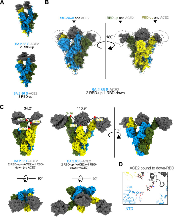
Fig. 2. Cryo-EM maps of BA.2.86-S-protein bound to ACE2.
A–C Cryo-EM maps of BA.2.86-S-protein bound to human ACE2 (S; same colors as Fig. 1C, ACE2; dark gray). A Two-RBD-up state (top), and three-RBD-up state (bottom). B Two-RBD-up−one-RBD-downthree-ACE2 state. C Comparison of the angles formed by the three residues: N164 of the NTD and two G476 of the up-RBD for two-RBD-uptwo-ACE2 (34.2°, left) and two-RBD-up−one-RBD-downthree-ACE2 (110.9°, right), respectively. D Close-up view of the two-RBD-up−one-RBD-downthree-ACE2 state. The N165-linked glycan in the NTD and the N322-linked glycan in ACE2 are situated close to each other.

