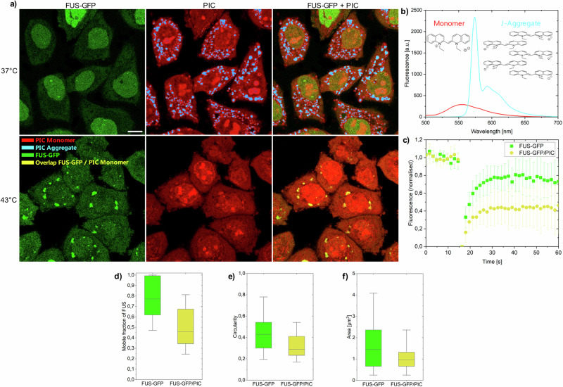
Fig. 1. PIC accumulation in HeLaFUS-GFP cells.
a HeLaFUS-GFP treated with PIC at 37 °C (upper images) and 43 °C (lower images). At 37 °C, FUS-GFP was mainly located in the nucleus and PIC aggregated in the cytoplasm. At 43 °C FUS-GFP accumulated in the cytoplasm and enriched in stress granules. PIC is monomeric in the cytoplasm and stress granules. J-aggregates (blue) and monomers (red) were assigned based on their intensity and spectrum (b) (see “Methods” section for details). Scale bar: 10 µm. c, d FRAP measurements of FUS-GFP in stress granules in presence and absence of PIC. e, f Circularity and area of stress granules in presence and absence of PIC. d–f Boxplot including the first quartile, median and third quartile of 346 individual cells. Whiskers represent 5–95% of all data points. Details in Supplementary Tables 1 and 2.

