
Fig. 4.
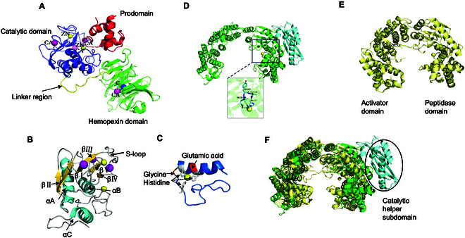
Collagenase crystal structures. (A) Ribbon representation of proMMP1. The MMP domains and inorganic ions are labeled and colored. (B) Ribbon representation of the catalytic site of proMMP1, with secondary structures colored and important segments labeled. (C) A ribbon representation of the active site of MMP1 is depicted, with the overall structure elegantly highlighted in a captivating teal hue. (D) Ribbon representation of ColG; the HEXXH zinc-binding motifs are labeled. (E) Ribbon representation of VhaC. (F) A comparison between the CM structures of VhaC (yellow) and ColG (green) is shown, and the catalytic helper subdomain of ColG is indicated by the circle.