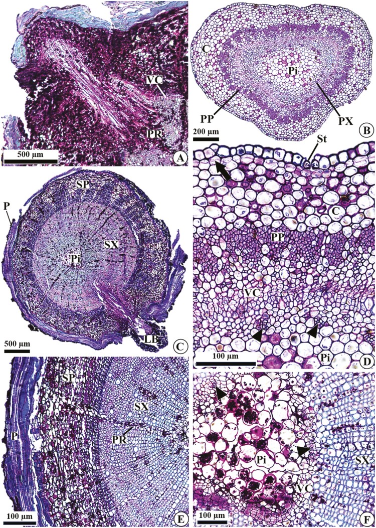
Figure 6.
Stem anatomy of D. filipedunculata in longitudinal (A) and cross (B–F) sections. (A) Part of the sprout connected to the root, associated with a wide parenchymatous ray. (B–C) General aspects in the apical and basal regions. (D) Details of the primary structure, with the establishment of phellogen below the epidermis. (E) Details of the secondary structure, with periderm, secondary phloem and secondary xylem. (F) Details of the pith in a stem with secondary growth. Arrow, phellogen; arrowheads, primary xylem; C, cortex; P, periderm; Pi, pith; PP, primary phloem; PR, parenchymatous ray; PX, primary xylem; SP, secondary phloem; St, stomata; SX, secondary xylem; VC, vascular cambium.

