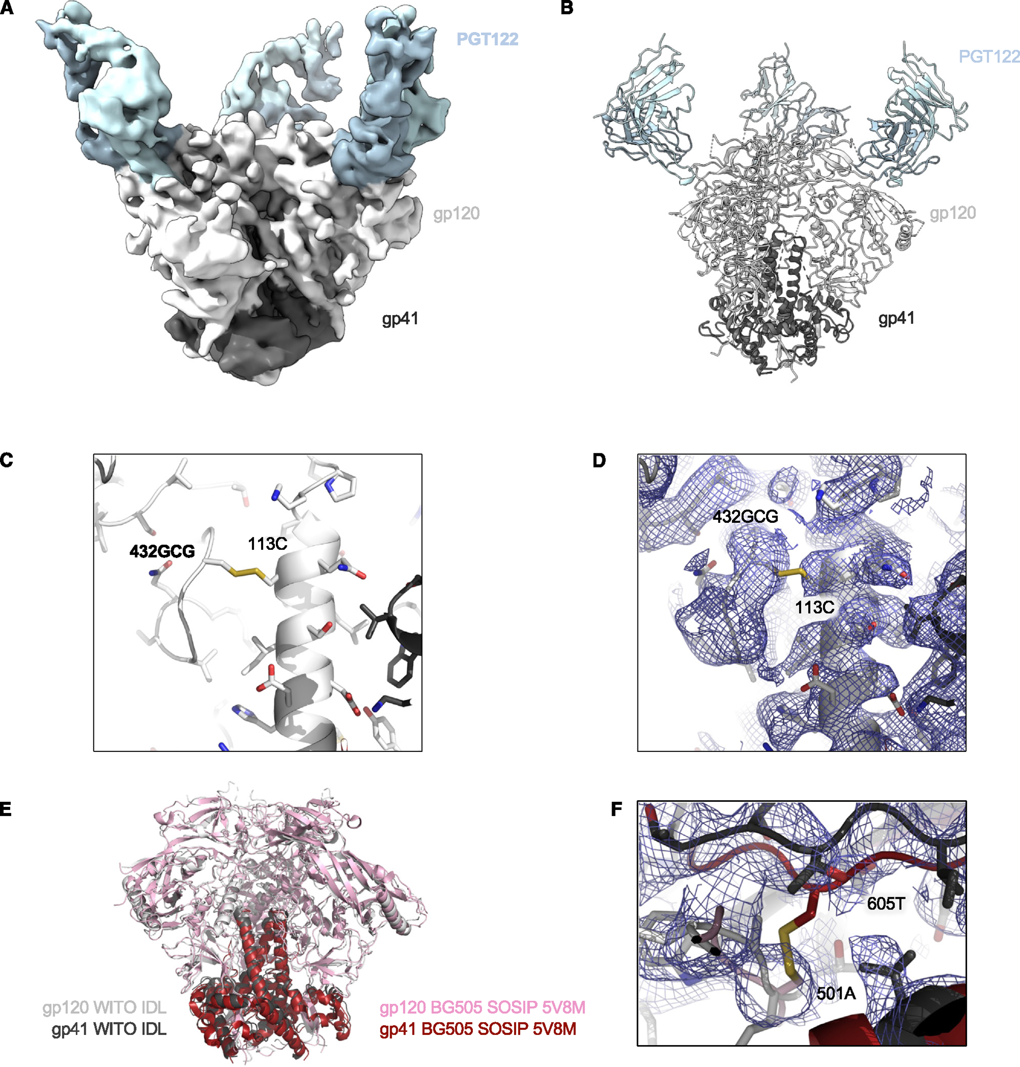
Figure 6. Cryo-EM structure of the HIV-1 WITO IDL Env trimer in complex with the Fab PGT122.

(A) Cryo-EM density is shown for HIV-1 Env WITO IDL bound to PGT122 Fab.
(B) Illustration of the trimer structure.
(C) Illustration and stick representation of the engineered disulfide bond.
(D) Volume density is shown in blue mesh and clearly resolved around the disulfide linkage shown in (C).
(E) Overall alignment of gp120 and gp41 IDL (gray and black) compared to BG505 SOSIP (pink and maroon, PDB: 5V8M). Illustrations of Env trimers are shown with bound Abs hidden.
(F) The region of disulfide stabilization in SOSIP trimers is shown contrasted with the structure we present colored as in (E), with density as blue mesh.
See also Figures S6 and S7 and Table S1.
