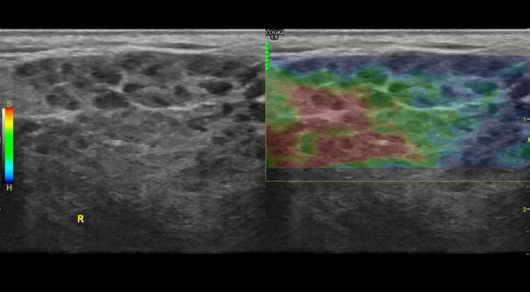
Figure 13.
Parotid gland: elastography of the right parotid gland from a patient with Sjögren’s syndrome. Ultrasound machine with 4D imaging. (GE Logic 9). Heterogeneous density with cystic areas and soft and compressible differentiated focal glandular images (S-red) and rigid non-compressible areas (H-blue).

 Content of this journal is licensed under a