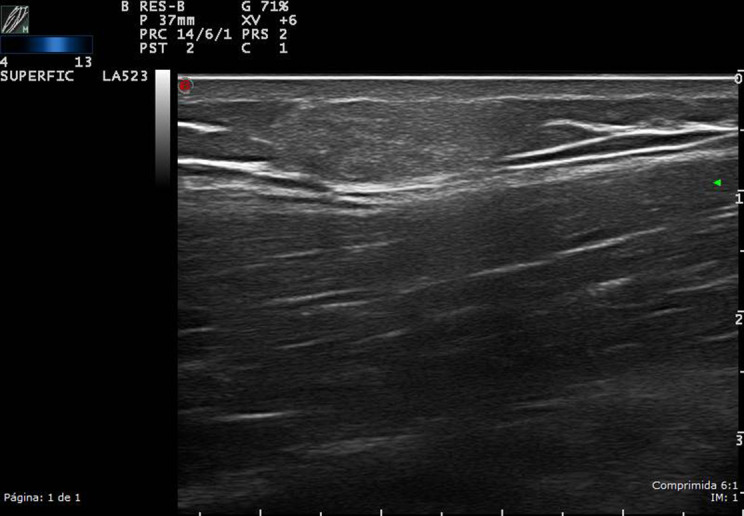
Figure 5.

High-resolution ultrasound lipoma of the right arm in grayscale, in a patient with a soft, slow-growing tumor in the proximal arm. The upper part of the arm was explored: a well-defined, homogeneous, isoechoic oval, superficial lesion of approximately 1.5 cm2.

 Content of this journal is licensed under a