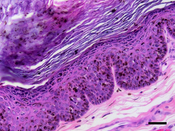
Figure 2.
Papillomaviral cutaneous plaques, dog. The plaque consists of heavily pigmented, hyperplastic epidermis covered by increased quantities of keratin. Large quantities of pigment and marked clumping of keratohyalin granules are visible within the thickened epidermis. Neither koilocytosis nor the presence of cells with expanded, blue-grey cytoplasm are visible within these lesions. PCR was used to amplify canine papillomavirus type 18 DNA from this plaque. Scale bar = 25μm. Haematoxylin and eosin.

