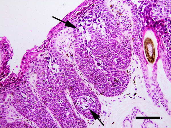
Figure 4.
Papillomaviral cutaneous plaque, cat. The epidermis is thickened and contains increased pigment. There is crowding of cells within the basilar layers. Papillomavirus-induced cellular changes are prominent including cells with expanded clear cytoplasm containing perinuclear bodies (arrows). PCR amplified Felis catus papillomavirus type 3 DNA from this plaque. Scale bar = 40μm. Haematoxylin and eosin.

