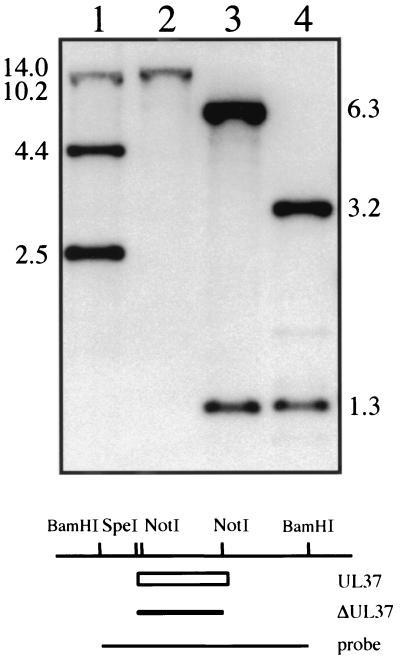
FIG. 2.
Southern blot analysis of the KΔUL37 genome. Two micrograms of KOS (lanes 1 and 3) and KΔUL37 (lanes 2 and 4) viral DNA was digested with NotI (lanes 1 and 2) or BamHI and SpeI (lanes 3 and 4) and resolved by agarose gel electrophoresis prior to analysis by Southern blot hybridization. Filters were probed with a 32P-labeled DNA probe corresponding to the BamHI H fragment. The size of hybridized fragments in kilobases is indicated at the sides of the gel. The schematic at the bottom of the figure shows the BamHI H region of HSV-1. The open box depicts the UL37 ORF, the filled box depicts the UL37 deletion, and the line at the bottom depicts the probe used for hybridization. Relevant restriction enzyme sites are shown at the top.

