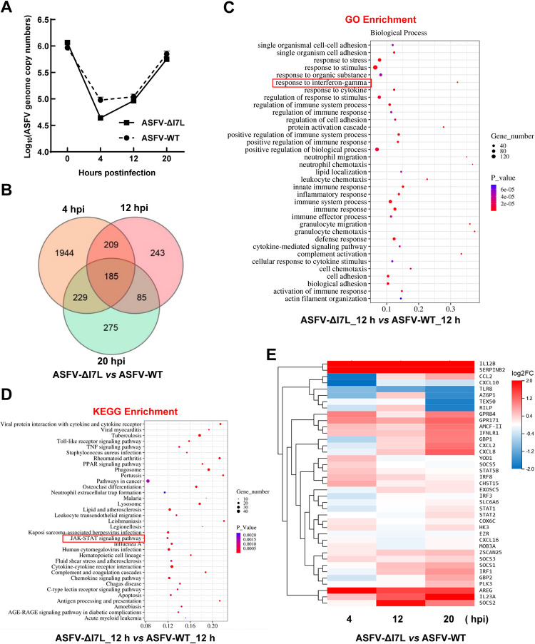
Fig 2. Differential expression profiling in the ASFV-ΔI7L-infected primary porcine alveolar macrophages (PAMs) by RNA sequencing analysis.
(A) Identification of ASFV infection. PAMs were infected with ASFV-ΔI7L or ASFV-WT at a multiplicity of infection (MOI) of 1. The ASFV genome copies were determined by a quantitative real-time PCR (qPCR) at 4, 12, and 20 hours postinfection (hpi). (B) Venn diagrams of the differentially expressed genes (DEGs) in the ASFV-ΔI7L- versus (vs.) ASFV-WT-infected PAMs. (C and D) The bioinformatics analysis of DEGs. The gene ontology (GO) enrichment (C) and the Kyoto Encyclopedia of Genes and Genomes (KEGG) enrichment (D) analyses were performed in the ASFV-ΔI7L- vs. ASFV-WT-infected PAMs at 12 hpi. (E) Heat map of the DEGs induced by ASFV-ΔI7L vs. ASFV-WT at 4, 12, and 20 hpi. Error bars denote the standard errors of the means. All the data were analyzed using the one-way ANOVA. ***, P < 0.001; ns, not significant.

