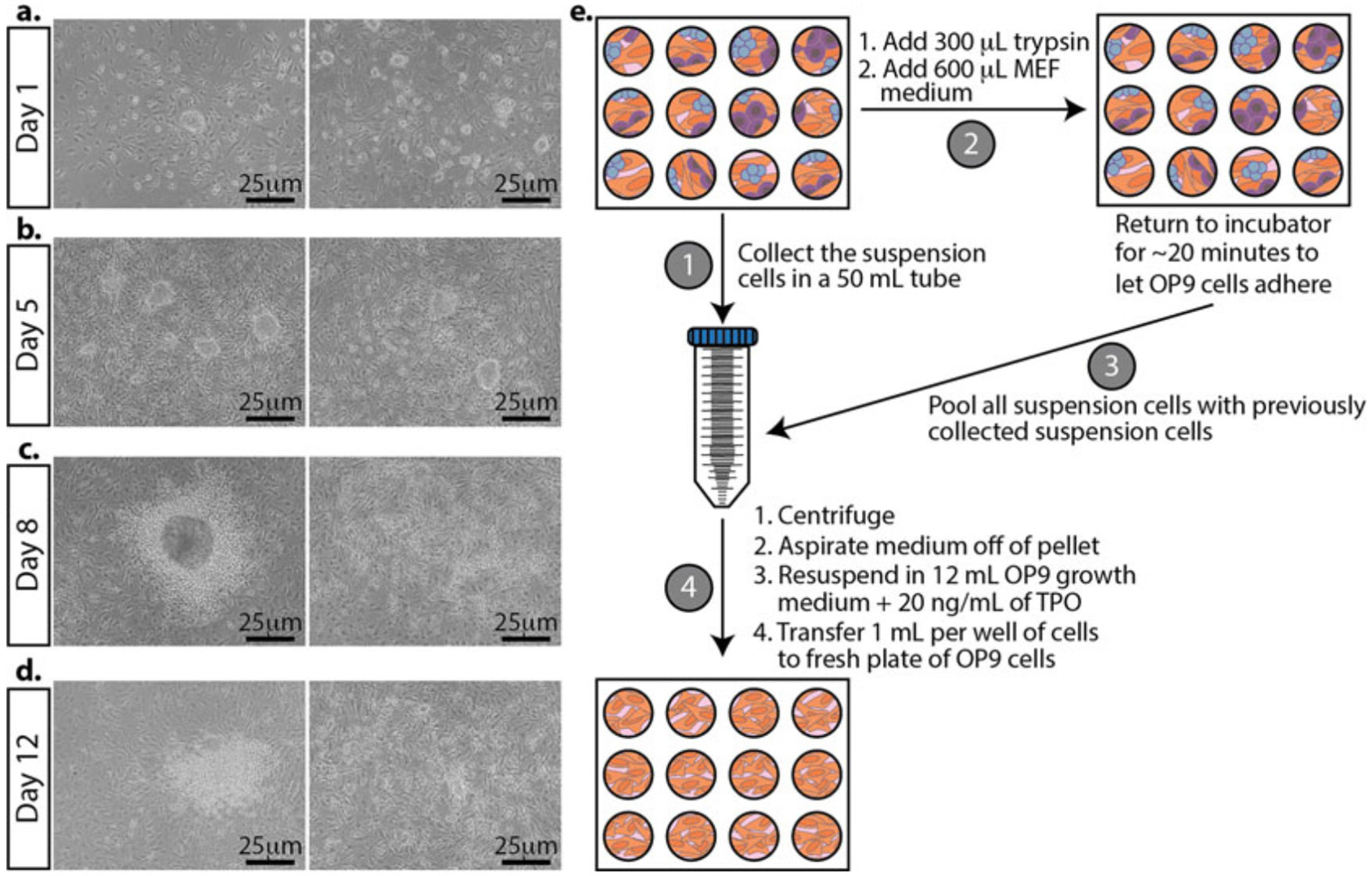
Fig. 5.

Morphological changes in mESCs during differentiation and passing differentiating mESCs. Representative bright-field images. (a) The day after plating mESCs on OP9 cells. (b) mESCs grown on OP9 cells for 5 days. TPO is added at this stage. (c) Three days after adding TPO to the cultures. (d) The last day of differentiation. The cells are ready to be analyzed for differentiation markers. (e) Schematic for differentiating mESCs. First, collect and pool the suspension cells from each well (left, labeled step 1). Second, add trypsin to the adherent cells. Once the cells are detached and the trypsin is neutralized with the medium, return the plate to the incubator to remove the majority of the OP9 cells (they will attach first) for about 20 min (top, labeled step 2). After the majority of the OP9 cells have attached, take the cells in suspension and pool them with the previously collected suspension cells (right, labeled step 3). Plate onto a fresh plate of OP9 cells growing in a 12-well plate (bottom, labeled step 4)
