. 2024 Oct 8;13:RP98070. doi: 10.7554/eLife.98070

Figure 5. Select DTX RING-DTC domains catalyse ubiquitination of ssDNA.

(A) Fluorescently detected SDS-PAGE gel of in vitro ubiquitination of 6-FAM-labelled ssDNA D4 by DTX3L-RD or DTX1-RD in the presence of E1, UBE2D2, Ub, Mg2+-ATP. (B) As in (A) but with DTX2-RD. (C) As in (A) but with DTX3-RD. (D) As in (A) but with DTX4-RD. (E) As in (A) but with DTX3L-RD or DTX3L RING with increasing concentrations of DTX3L DTC. (F) Fluorescently detected SDS-PAGE gel of in vitro ubiquitination of 6-FAM-labelled ssDNA D1-9 by DTX2-RD. A reaction with DTX3L-RD and 6-FAM-labelled ssDNA D4 was included as a positive control. (G) Western blot of in vitro ubiquitination of biotin-NAD+ in the presence of E1, UBE2D2, Ub, Mg2+-ATP, NAD+, biotin-NAD+ with either DTX3L-RD, DTX3LR-DTX2D, or DTX2R-DTX3LD and separated by SDS-PAGE. (H) Fluorescently detected SDS-PAGE gel of in vitro ubiquitination of 6-FAM-labelled ssDNA D4 by DTX3L-RD, DTX3LR-DTX2D, or DTX2R-DTX3LD in the presence of E1, UBE2D2, Ub, Mg2+-ATP. (I) Schematic diagrams showing the proposed mechanism of ubiquitination of substrates by DTX3L-RD. Asterisks in (A–F) and (H) indicate contaminant bands from ssDNA. Raw unedited and uncropped gel images of (A–H) are shown in Figure 5—source data 1 and 2, respectively.

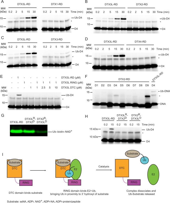
Figure 5—source data 1. Raw unedited gels for Figure 5.
Figure 5—source data 2. Uncropped and labelled gels for Figure 5.

Figure 5.

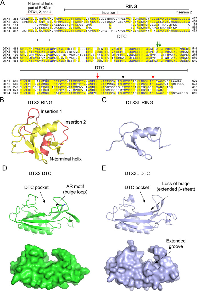
Figure 5—figure supplement 1. Properties of DELTEX (DTX) family DELTEX C-terminal (DTC) domains.

Figure 5—figure supplement 1.

(A) Sequence alignment of human DTX family RING-DTC domains. Starting and ending residues of RING-DTC domains are indicated. Conserved residues are highlighted in yellow. A black arrow indicates residue that stacks with adenosine 5′-diphosphate (ADP)–ribose (ADPr), red arrows indicate conserved catalytic residues, and green arrows indicate AR motif present in DTX1, 2, and 4. Insertions are indicated. (B) Structure of DTX2 RING domain (from PDB: 6Y3J). The insertions and N-terminal helix are colored in red and the conserved RING domain region is colored yellow. (C) Alphafold2 model of DTX3L RING domain (light blue) displayed in the same orientation as in B. (D) Top: cartoon representation of the structure of DTX2 DTC domain (green; PDB: 6Y3J). Sidechains of the AR motif are shown as sticks. The DTC pocket that binds ADPr and the bulged AR motif are indicated by arrows. Bottom: as in the top panel but with surface representation. (E) Top: cartoon representation of the structure of DTX3L DTC domain (light blue; PDB: 3PG6). Bottom: as in the top panel but with surface representation. The absence of an AR motif in DTX3L causes a slight structural change near the ADPr-binding pocket, leading to the loss of the bulged loop, which results in a slightly extended groove.