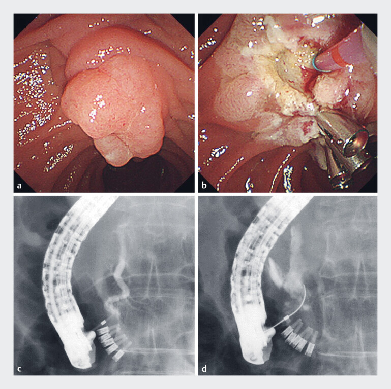
Fig. 1.
Images during endoscopic papillectomy showing: a, b on endoscopic view: a an ampullary tumor that was localized to the papilla; b pancreatic duct cannulation being attempted after prophylactic clipping had been carried out; c, d on fluoroscopic view; c significant bends within the main pancreatic duct; d pancreatic duct injury caused by guidewire penetration into the retroperitoneal cavity.

