Fig. 4.
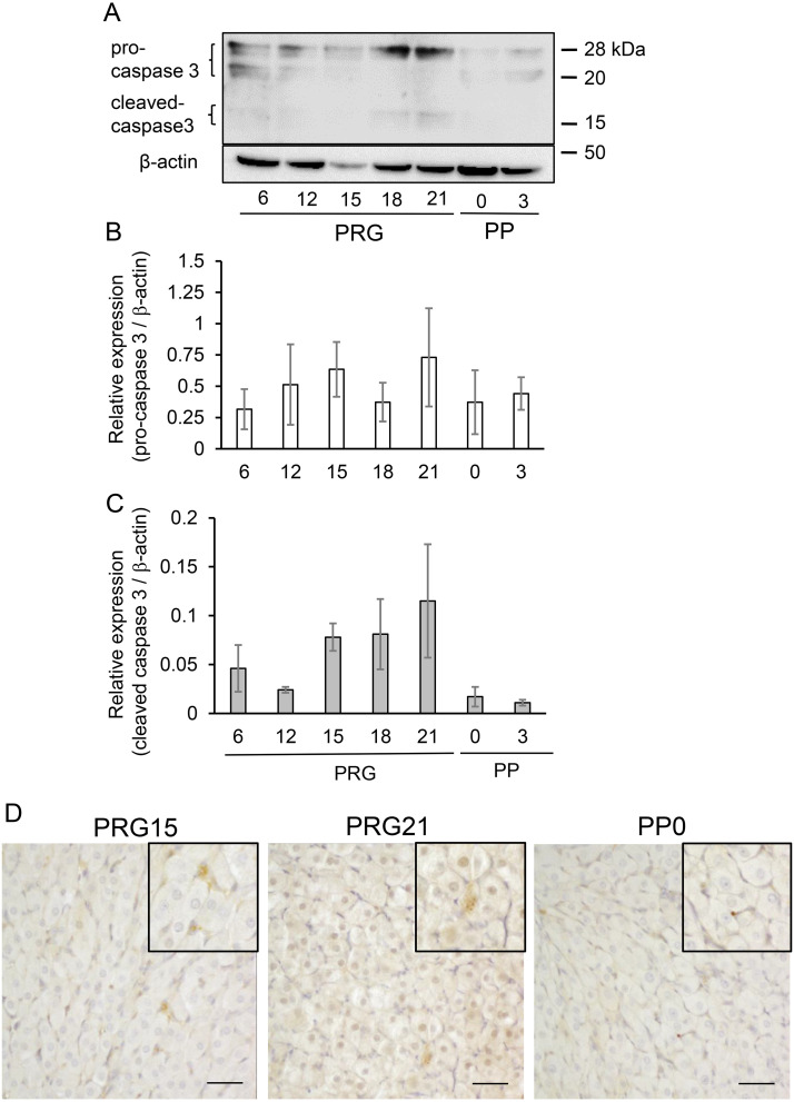
Expression of caspase 3 protein in CL tissue. (A) Typical immunoblots of caspase 3 in CL tissue throughout pregnancy and postpartum. Protein expression of pro-caspase 3 (B) and cleaved caspase 3 (C) relative to β-actin was determined and shown (mean ± SEM, n = 3). (D) Immunostaining of cleaved caspase 3 in CL. Note the enhanced positive reaction in luteal cells on PRG21 when compared to those on PRG15 and PP0. Scale bar, 50 µm.

