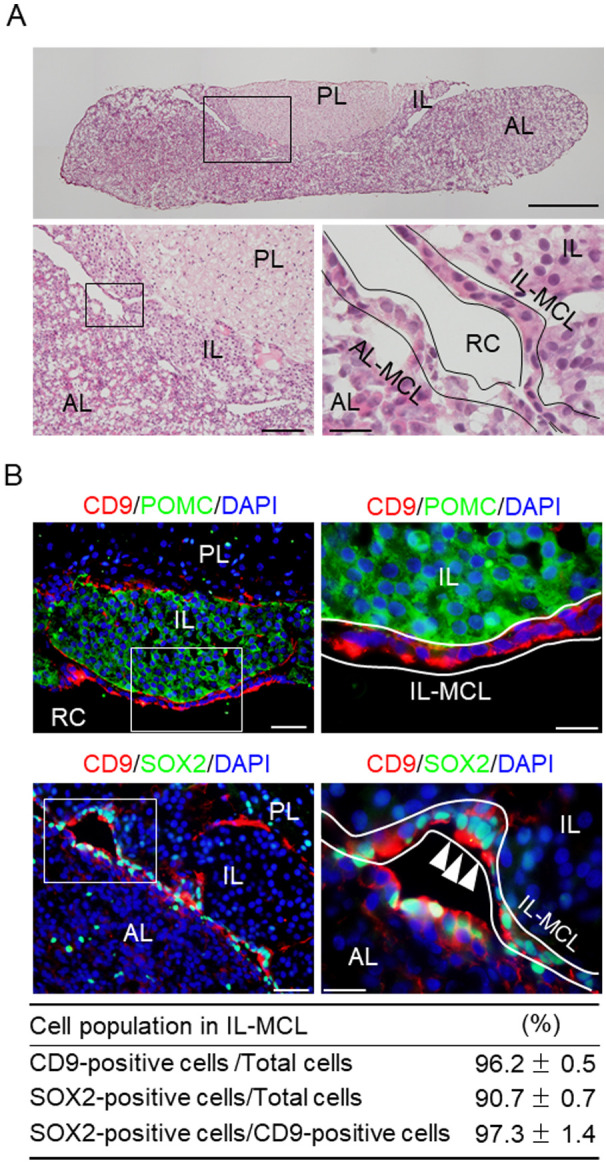
Fig. 1.

CD9-positive cells in the marginal cell layer (MCL) of the intermediated lobe (IL) in the rat pituitary gland. (A) Hematoxylin and eosin staining of a cryosection of the pituitary gland of a male adult rat. Lower panels: Higher magnification of the boxed area in the upper (left) and left panels (right). (B) Merged images of 4’,6-diamidino-2-phenylindole (DAPI) staining (blue) and double immunofluorescence staining for CD9 (red) and POMC (upper panels, green) or SOX2 (lower panels, green). The right panels show high-magnification images of the boxed areas in the left panels. White arrowheads indicate CD9/SOX2-positive cells. Proportions of CD9-or SOX2-positive cells among DAPI-positive cells or SOX2-positive cells among CD9-positive cells in a rectangular area (157.5 × 210 μm) in the MCL of the IL are shown in the bottom table (mean ± SEM, n = 5). PL, posterior lobe. AL-MCL, MCL of AL. IL-MCL, MCL of IL. RC, Rathke’s cleft. Scale bars: 500 μm (upper panel of A), 100 μm (lower left panel of A), 50 μm (lower right panel of A and upper and lower left panels of B), and 20 μm (lower right panel of A, upper and lower right panels of B).
