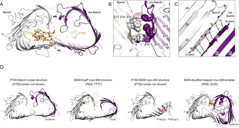
Fig. 4. Crystal structure of the open lateral gate PTB2–BamA complex.
A Overall arrangement of two PTB2–BamA complexes observed within the X-ray crystal lattice. One BamA molecule is named ins-BamA (purple), and the other BamA is shown in gray. The PTB2 macrocycle is shown in orange. Positions of the views shown in panels B and C are noted with the corresponding letter. B Close-in view of the BamA (gray)/ins-BamA (purple) β14–β16/β14–β16 interaction interface. C Close-in view of the BamA (gray)-ins-BamA (purple) β1–β1 interaction interface. D Comparison of the crystal structure of PTB2-BamA (left, BamA in gray and ins-BamA in purple), the cryo-EM structure of BAM–EspP complex8 (middle-left, BamA in gray and EspP in purple), the cryo-EM structure of PTB2-BAM (middle-right, BamA in gray, PTB2-lg-1 shown in pink and PTB2-lg-2 shown in green), and the cryo-EM structure of BAM–disulfide trapped complex7 (right, BamA in gray, subBamA in purple, and disulfide in yellow).

