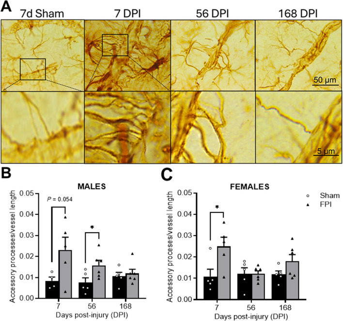
FIGURE 3.
TBI promotes time-dependent changes in the number of GFAP-labeled accessory processes. (A) Representative GFAP immunostained images within the S1BF region of sham and FPI rats at 7, 56, and 168 DPI from both sexes to highlight changes in accessory processes over time. The top row are images captured at 100×. The bottom row images magnify the accessory process interactions with the vessel. (B) In males, an FPI increased accessory processes at 56 DPI compared to age- and sex-matched shams. (C) In females, FPI increased primary processes at 7 DPI compared to sham N=5–6/sex. Data were analyzed by a two-way ANOVA followed by Tukey’s multiple comparison tests. *P < 0.05 and **P < 0.01. Error bars indicate + SEM. Scale bar = 50 µm.

