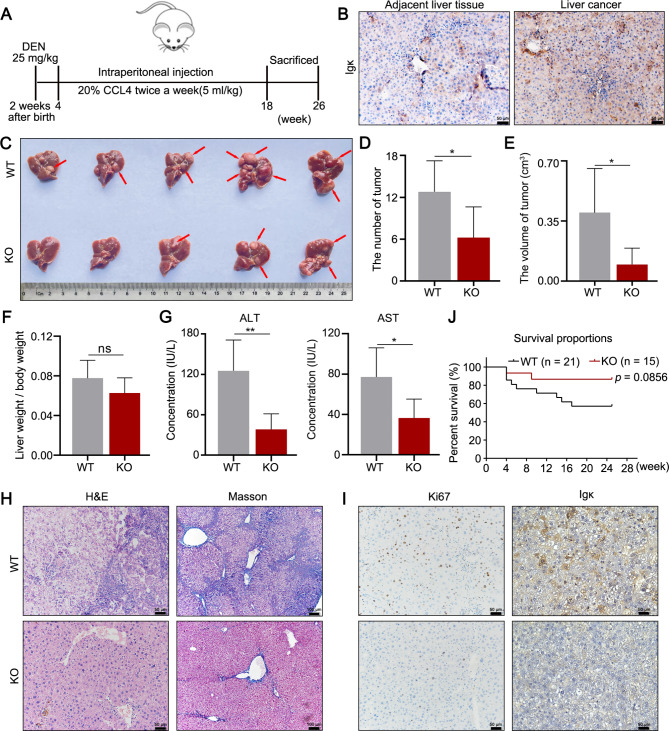
Fig. 3.
Hepatocyte-specific depletion of Igκ suppresses HCC progression in mice. A Schematic representation outlining the procedure for DEN and CCL4-induced HCC mouse models. B Representative images of immunohistochemical staining for Igκ protein expression in liver cancer tissues and adjacent liver tissues from mice with HCC. C Tumor formation in liver tissues was compared between WT and KO mice, and the red arrow indicates typical tumor nodes (n = 5). D - F Statistical analysis of the tumor number (D), maximum tumor volume (E) and ratio of liver weight to body weight (F) (n = 5). G The levels of serum ALT and AST were measured. H H&E and Masson staining were performed to assess the pathological changes in the liver tissues of WT and KO mice (n = 5). Scale bars, 50 μm and 100 μm. I The expression of Ki67 and Igκ in liver tissues was determined by immunohistochemical staining. Scale bar, 50 μm. J Survival analysis of WT and KO mice with DEN and CCL4-induced HCC. The data are presented as the mean ± SD. * p < 0.05, ** p < 0.01

