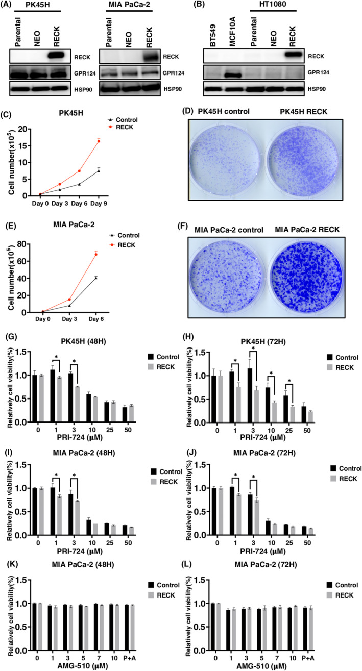
FIGURE 2.

RECK stimulates cell proliferation in a WNT signal‐dependent manner when induced in GPR124‐positive pancreatic ductal adenocarcinoma (PDAC) cells. (A, B) Immunoblotting (IB) of the indicated proteins in the indicated cells parental or transduced with PCXN2‐NEO or PCXN2‐RECK. (C) Cell proliferation of PK45H transduced with PCXN2‐NEO or PCXN2‐RECK (N = 3). (D) Representative image of colony formation by PK45H cells transduced with PCXN2‐NEO or PCXN2‐RECK (N = 3). (E) Cell proliferation of MIA PaCa‐2 cells transduced with PCXN2‐NEO or PCXN2‐RECK (N = 3). (F) Representative image of colony formation by MIA PaCa‐2 transduced with PCXN2‐NEO or PCXN2‐RECK (N = 3). (G–L) Relative viability of the indicated cells transduced with PCXN2‐NEO or PCXN2‐RECK and treated with the indicated concentration of the indicated chemicals for the indicated period. The viability of cells without treatment was set to 100% (N = 3). *p < 0.05 against vector control; N.S., not significant.
