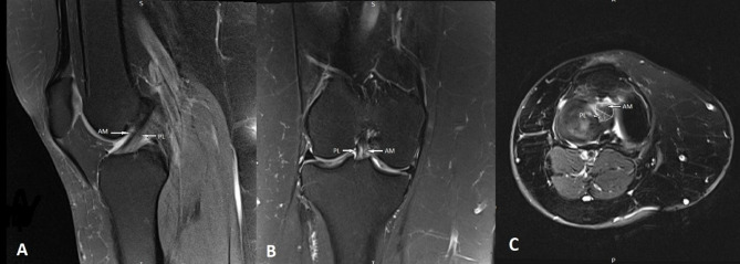
Figure 1.
Normal anterior cruciate ligament anatomy in which the AM and PL bundles are clearly delineated from each other; (A) sagittal proton density (PD) weighted fat suppressed image demonstrating the normal ACL, characterized by taut, continuous, low intensity ACL fibers, (B) coronal T2 weighted fat suppressed image, and (C) axial T2 weighted fat suppressed knee MRI image that shows the AM and PL footprint.

