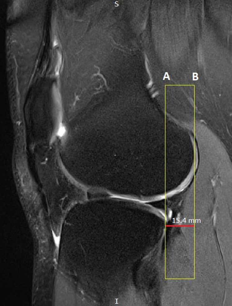
Figure 5.
Proton density weighted fat suppressed sagittal image demonstrates anterior tibial translation in association with ACL injury; line (A) is crossing the most posterior point of the posterolateral tibial plateau and is parallel to line (B) which is crossing the most posterior point of the lateral femoral condyle. The distance between these two lines is 15.4mm, indicating an ACL tear of the knee.

