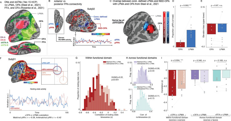
Fig. 3.
Retinotopic coding structures the spontaneous opponent interaction between functionally-coupled mnemonic and perceptual areas during resting-state fMRI. A. Isolating functionally coupled internally- and externally-oriented brain areas within the DNs and dATNs. We established brain areas relevant to two cognitive domains, visual analysis of 1) scenes and 2) faces. Specifically, we focused on a memory area in the domain of scene perception (the lateral place memory area (LPMA; from43), white) at the posterior edge of the DN-A (purple; from33). We examined LPMA’s relationship to a set of perceptual areas in the dATN (green; from33), 1) the occipital place area (OPA; from43), an area within the domain of scene perception, along with 2) the occipital face area (OFA) and 3) the fusiform face area (FFA), two areas involved in the domain of face perception (white; from51). B-C. We localized LPMA in all participants by contrasting the correlation in resting-state activity between anterior and posterior parahippocampal place area (PPA) (B). This yielded a region in lateral occipital-parietal cortex that overlapped with the LPMA defined in an independent group of participants (C). D-E. Consistent with prior work, the connectivity-defined LPMA had greater concentration of −pRFs compared to OPA (D), and exhibited a lower visual field bias to OPA (E), consistent with an opponent interaction between these areas during perception. F. We assessed the influence of retinotopic coding on the interaction between −pRFs in mnemonic and +pRFs in perceptual areas using the same pRF matching and correlation procedure described above. We compared pRFs within functional domain (scene memory × perception –LPMA to OPA) as well as across domains (scene memory × face perception –LPMA to the occipital face area (OFA) and fusiform face area (FFA)). G. Within functional domain opponent interaction reflects voxel-wise retinotopic coding. We observed a stronger negative correlation between matched compared to anti-matched −LPMA/+OPA pRFs (-LPMA × +OPA matched versus anti-matched pRFs: D(392)=0.22, p<0.001). H. Retinotopic coding did not impact the interaction between areas across functional domains. We found no significant difference between matched and anti-matched pRFs between the scene memory area LPMA and the face perception areas FFA and OFA (-LPMA × +OFA: D(392)=0.09, p=0.44; −LPMA × +FFA: D(392)=0.11, p=0.15). Histograms depict the distribution of correlation values between matched (dark) and antimatched (light) pRFs for all runs in all participants. I. Retinotopic coding organizes interactions within a domain, but not across domains. When the correlation values were averaged within each participant, we observed a significant difference between matched versus anti-matched pRFs within functional domain (-LPMA × +OPA: t(6)=4.45, p=0.004) but not across domains (-LPMA × +OFA: t(6)=1.04, p=0.34; −LPMA x +FFA: t(6)=1.48, p=0.188).

