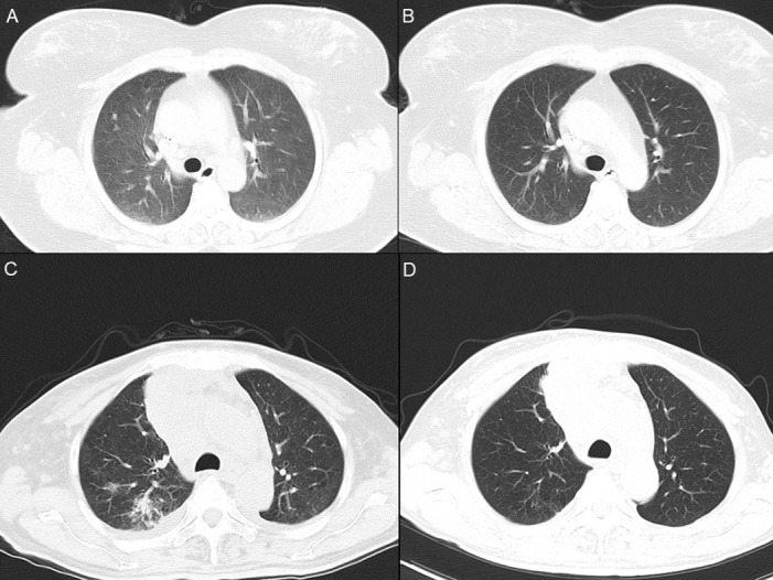
Fig 1. Thoracic computed tomography scan of Case 2 and 3 before and after specific antibiotic treatment.

Panel A: Case 2, day 9 after admission, showing diffuse interstitial infiltrates and ground-glass nodules in both lungs; Panel B: Case 2, day 40 after admission, showing clearance of infiltrates and nodules; Panel C: Case 3, day 5 after admission, showing infiltrates in the posterior segment of the upper lobe of the right lung; Panel D: Case 3, day 64 after admission, showing clearance infiltrates.
