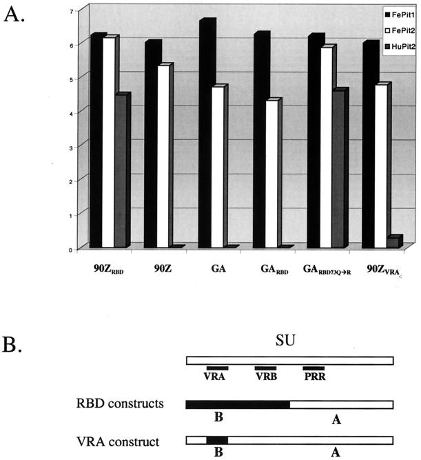
FIG. 2.
Infection of cells expressing Pit1 or Pit2 by viruses pseudotyped with various FeLV-B SUs. (A) Single-cycle infection assay that results in the transduction of a vector genome encoding β-galactosidase. The x axis indicates the envelope-SU of the infecting pseudotyped virus, and the y axis indicates the focus-forming units (FFU) per milliliter of viral supernatant as determined by β-galactosidase activity in a log scale. Within the graph, the shading of the bar depicts the target cells for infection. As shown in the key at the top of the graph, solid black bars, white bars, and gray bars indicate MDTF-FePit1s, MDTF-FePit2s, and MDTF-HuPit2s, respectively. Naive MDTF cells were also exposed to virus in parallel, and no background was observed (55) (data not shown), except in the case of FeLV-B-90ZRBD, which gave an average of 20 FFU/ml in five replicate experiments using the same virus stocks. MDTFs expressing HuPit1 were also infected in parallel, and the results were always similar to those obtained with MDTF-FePit1 cells (55) (data not shown). (B) Schematic of the chimeric FeLV envelope-SUs tested in panel A. The small black boxes show the relative positions of the VRA, VRB, and PRR regions in the SU. The schematic depicting the RBD constructs (GARBD, GARBD-73Q→R and 90ZRBD) illustrates that these pseudotyped viral SUs contain regions derived from FeLV-B (in black) that include the VRA and VRB regions, while the rest of the SU, including the PRR, is supplied by FeLV-A (in white). In the VRA construct, only the VRA region is derived from FeLV-B, while the remainder of the SU is FeLV-A derived.

