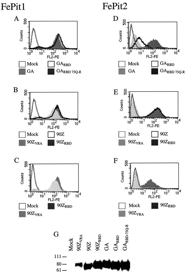
FIG. 3.
Binding of FeLV-B SU to MDTF cells expressing either FePit1 or FePit2. Panels A to F show overlays of flow cytometry data of FeLV SU-HA supernatants that had been bound to either MDTF-FePit1 cells (panels A, B, and C) or MDTF-FePit2 cells (D, E, and F). Binding was detected using an HA monoclonal antibody. The x axis is fluorescence intensity (log scale), and the y axis is cell number. In panels A-F, the legend below each histogram indicates which FeLV SU-HA was used in the binding assay. In panels A to F, mock represents cells incubated with medium only. In panels A, B, D, and E, 1 ml of cell supernatant was used in the binding experiment. In panels C and F, 1 ml of FeLV-B-90ZVRA and 0.1 ml of FeLV-B-90ZRBD supernatant were used in order to compare more similar levels of protein (see panel G). Panel G is a Western blot that was performed with the supernatants used in the binding assays shown above. Equal amounts of supernatant were used. The sizes of markers (in kilodaltons) are indicated to the left of the blot. The methods used in the immunoprecipitation of the supernatants and Western blot procedure have been described previously (25).

