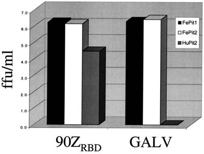
FIG. 4.
Infection of MDTF-FePit2 cells by GALV. The layout for infection studies is as described in the legend for Fig. 2A, where the targets cells are indicated in the upper right-hand corner and the viral pseudotypes used for infection are indicated on the x axis.

