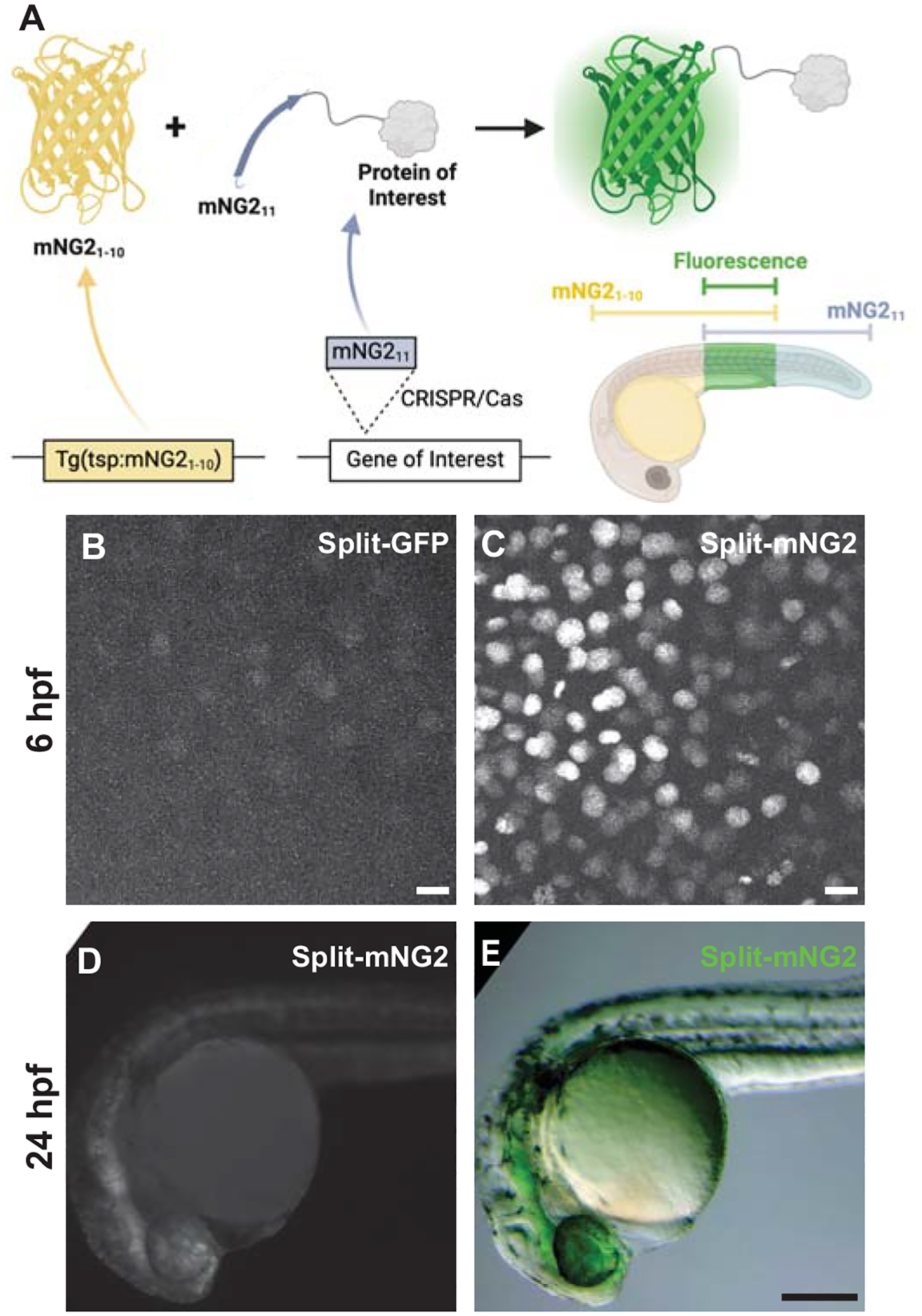
Figure 1. Split fluorescent protein fragments are functional in zebrafish embryos.

A. Schematic illustrating our protein labeling strategy using a split fluorescent protein. Transgenic (Tg) mNG21–10 is expressed under the control of a tissue-specific promoter (tsp) while mNG211 is inserted into protein-coding genes by CRISPR/Cas-directed gene editing. Fluorescence (green) is only generated in tissues co-expressing mNG21–10 and the mNG211-tagged protein of interest. B–E. Embryos were injected with GFP1–10 and GFP11-H2B (split-GFP, B) or mNG21–10 and mNG211-H2B (split-mNG2, C–E) mRNAs then imaged at 6 hours post-fertilization (hpf) on a confocal microscope (B, C) or at 24 hpf on a fluorescence stereomicroscope (D, E). Confocal images are displayed as maximum z-projections. Scale bars in B and C, 50 μm. Scale bar in E, 200 μm.
