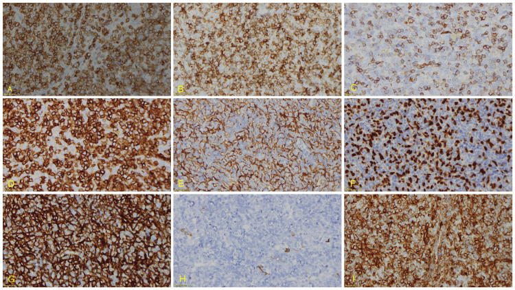
Figure 5. Immunohistochemistry panel of the nodule with the following stains: (A) CD3, (B) CD5, (C) CD45, (D) CD99, (E) CK 5/6, (F) p63, (G) CK (AE1/AE3), (H) CD20, and (I) vimentin.
Immunohistochemistry of the nodule showed positivity with the following stains: (A) CD3 (polyclonal), (B) CD5 (4C7), (D) CD99 (12E7), (E) CK 5/6 (D5/16 B4), (F) p63 (Dak-p63), (G) CK (AE1/AE3), and (H) CD20 (L26), while it showed patchy positivity with (I) vimentin (V9)

