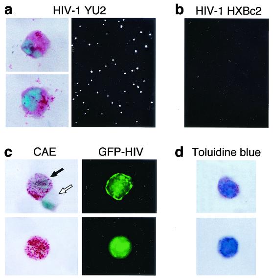
FIG. 3.
In situ detection of HIV in hPrMC and hMC. Purified hPrMC (4 weeks old) were infected with GFP-encoding viruses carrying envelope glycoproteins from the M-tropic HIV-1 YU2 (a) or the T-tropic HIV-1 HXBc2 (b). GFP-positive cells were then visualized by fluorescence microscopy. Up to 1% of the cells infected with the HIV-1 YU2 single-round virus showed green fluorescence (as depicted in panel a). No green cells resulted from infection with a similar virus carrying the envelope glycoprotein of HIV-1 HXBc2 (b). The GFP-HIV-positive cells sorted 5 days after exposure to HIV-1 YU2 were positive for CAE (a, left). Results are representative of three experiments performed. (c) When sorting was carried out 2 weeks after infection, the GFP-HIV-positive cells had strongly CAE-positive secretory granules (left, black arrow) and the few GFP-negative cells were also CAE negative (left, white arrow). (d) The granules of the sorted GFP-HIV-positive cells were also toluidine blue positive.

