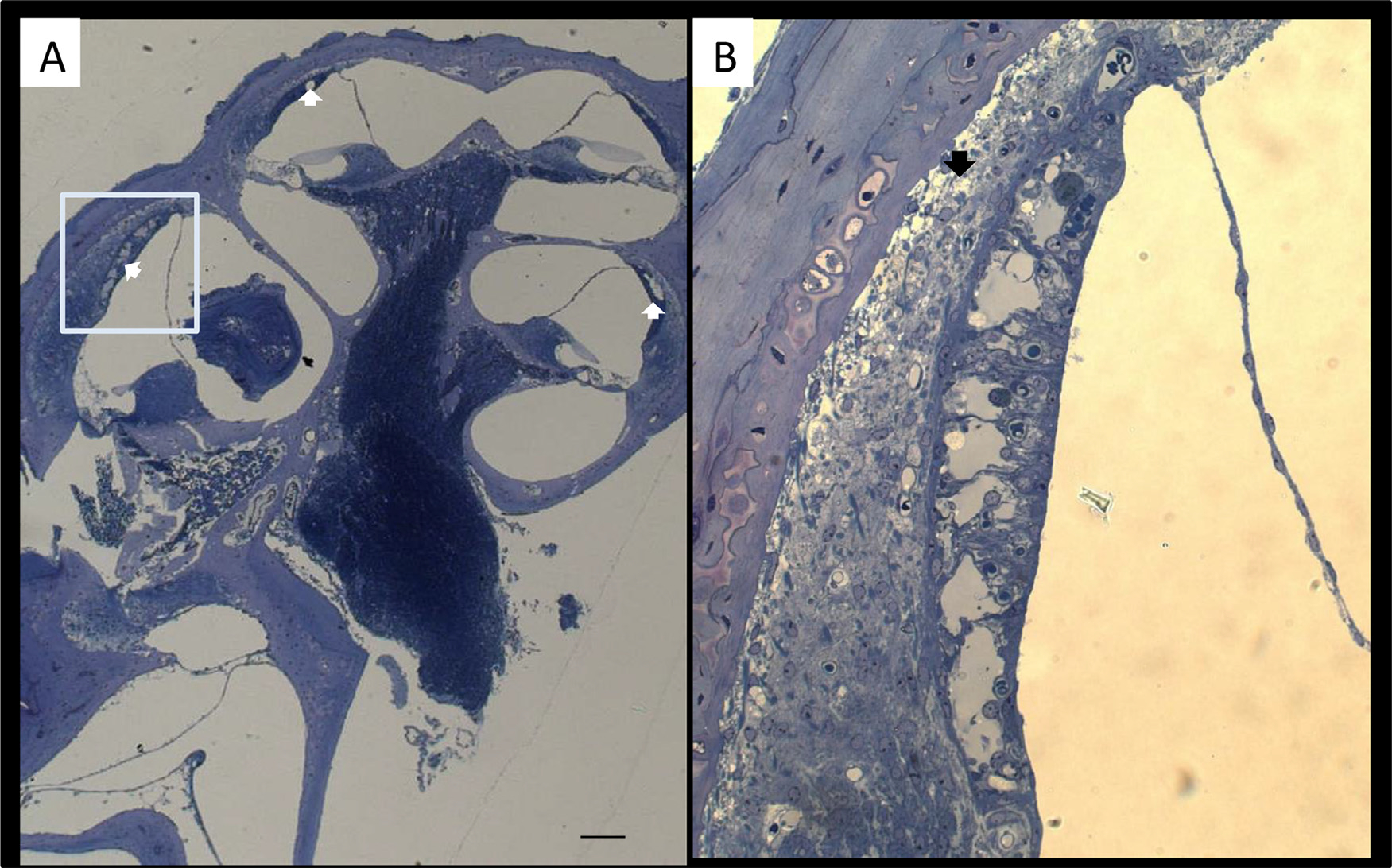
Fig. 2.

Representative light micrographs of the cochlea after administration of GeO2 for 4 months. Enlarged image of stria vascularis for captured area (B). Degeneration of the stria vascularis indicated with white arrow is seen in almost all cochlear turns, more markedly in the lower turn (A). Severe vacuolar degeneration of the stria vascularis is found in the lower basal turn (B). Bar = 100 μm in (A), 50 μm in (B).
