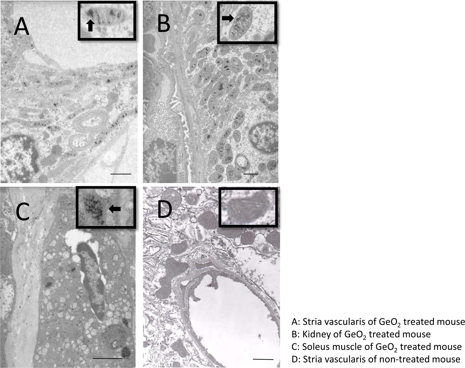
Fig. 3.

Ultrastructural findings of the intermediate cells of stria vascularis (A), kidney (B), soleus muscle(C) of GeO2 treated mice. Transmission electron microscope showed vacuolar degeneration of the stria vascularis (A). The arrow head indicates degenerated mitochondria containing electron-dense inclusion. The distal tubular epithelium of the kidney (B) and soleus muscles (C) showed many electron-dense deposits inside the degenerated mitochondria. Ultrastructural findings from a stria vascularis of non-treated mice (D).Inset: high-power view of mitochondria. andBar = 1μm.
