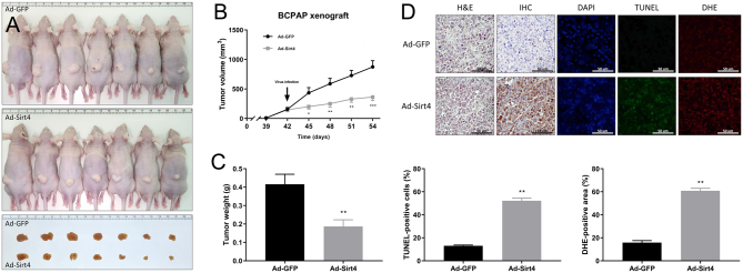
Figure 5.
SIRT4 inhibits tumor growth in a B-CPAP xenograft mouse model. (A) On day 42 after implanting tumor cells, mice received intratumoral injections of Ad-GFP or Ad-SIRT4. The tumor images show excised tumors from each group, with the top group representing the control group and the bottom group representing the SIRT4-expression group. (B) Tumor diameter was measured every 3 days using digital calipers and tumor volume was calculated. The diagram shows the tumor growth curve of the 14 mice at the indicated times after intratumoral injections of Ad-GFP or Ad-SIRT4. (C) Tumor weight was compared between the SIRT4-expression group and the control group. (D) SIRT4 overexpression induces apoptosis and superoxide production in mouse tumors. FFPE sections of tumors were co-stained with DAPI (0.5 µg/mL) to visualize nuclei, along with TdT enzyme (200 µg/mL) and DHE (10 μmol/L) to detect apoptosis and superoxide production, respectively. Apoptotic green fluorescence, oxidative red fluorescence, and nuclear blue fluorescence were analyzed under a fluorescence microscope at 400× magnification. The graph below shows the quantified signal of cells stained positively by TdT and DHE in five random fields. n = 8. *P < 0.05, **P < 0.01, ***P < 0.001. DAPI, 4′,6-diamidino-2-phenylindole; DHE, dihydroethidium; FFPE, formalin-fixed paraffin-embedded; TdT, terminal deoxynucleotidyl transferase; TUNEL, TdT-mediated dUTP nick end labeling.

 This work is licensed under a