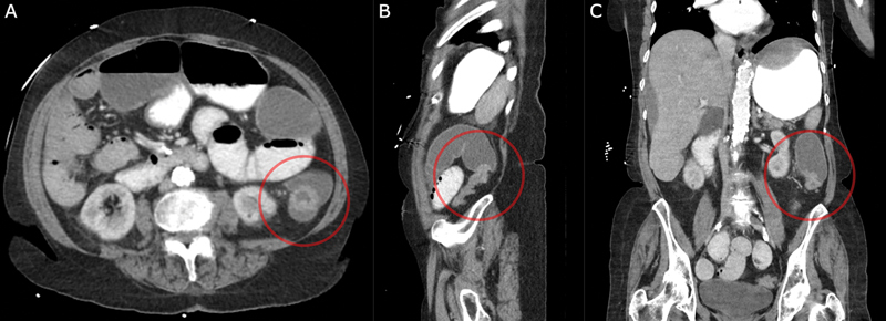
Fig. 1.

Computed tomography (CT) scan showing large bowel obstruction with dilated transverse colon and transition point in descending colon. ( A ) Axial views with a circle around the obstructing mass. ( B ) Sagittal views with a circle around the transition point. ( C ) Coronal views with a circle around the transition point.
