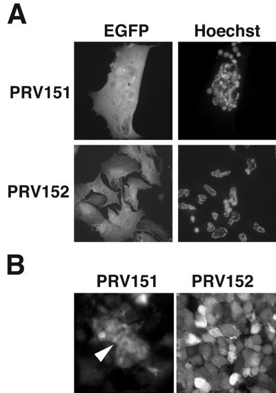
FIG. 1.
PRV infection of embryonic chicken retinal cells. (A) Infection of primary retinal cultures. Mixed retinal cultures 24 h after infection with PRV151 and PRV152. The EGFP signal is shown on the left, and the same cells stained with Hoechst 33258 are shown on the right. (B) Optical sections midway through PRV-infected chicken embryo retinal whole mounts 48 h after intravitreal infection with PRV151 (left) or PRV152 (right). The EGFP signal was visualized by confocal fluorescence microscopy. The arrowhead indicates fused cells.

