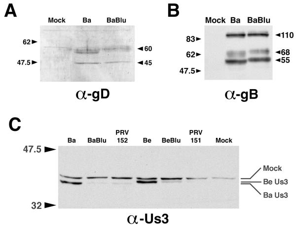
FIG. 7.
Reduced Us3 expression in cells infected with PRV strains that have insertions in gG. (A) Western blot of proteins extracted from MDBK cells (Mock) or MDBK cells infected with Bartha (Ba) or Bartha-Blu (BaBlu) probed with rabbit polyclonal antisera to PRV gD. (B) Western blot of proteins extracted from MDBK cells (Mock) or MDBK cells infected with Bartha (Ba) or Bartha-Blu (BaBlu) probed with goat polyclonal antisera to PRV gB. (C) Western blot of proteins extracted from MDBK cells (Mock) or MDBK cells infected with Bartha (Ba), Bartha-Blu (BaBlu), PRV152, Becker (Be), Becker-Blu (BeBlu), or PRV151, probed with an affinity-purified rabbit polyclonal antiserum raised against a synthetic peptide corresponding to the carboxy terminus of Us3. All infected cell extracts were prepared at 6 h postinfection. The positions of protein size markers (in kilodaltons) are shown to the left of the gels.

