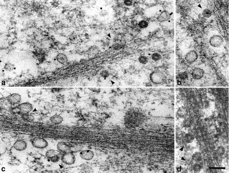
FIG. 6.
Movement of vesicles containing virions (a and b) or VP1 pseudocapsids (c and d) and empty vesicles derived from caveolae along actin filaments in NIH 3T6 fibroblasts. Cell sections were visualized by electron and immunoelectron microscopy. Staining was done with the rabbit anti-caveolin-1 polyclonal antibody followed by the 10-nm gold-goat anti-rabbit IgG antibody. Arrowheads point to vesicles containing viral particles. Bars, 100 nm.

