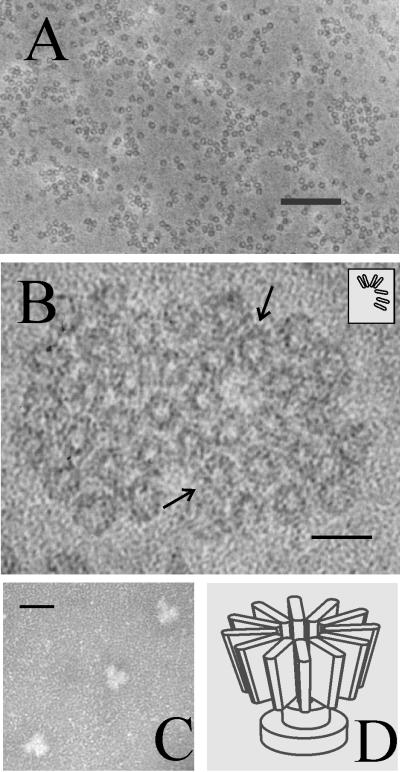
FIG. 5.
Electron microscopy of purified pUL6. Specimens were prepared by negative staining on standard (A and B) or glow-discharged (C) grids as described in Materials and Methods. Note that on standard grids, pUL6 is found in the form of small rings (A) and that at higher magnification the rings can be seen to be composed of subunits (B; arrows). The inset in panel B shows a schematic representation of the subunits present in the upper-arrowed ring. When adsorbed to glow-discharged grids, portal complexes were found to yield Y-shaped images, as shown in panel C. Shown in panel D is a diagrammatic representation of the portal complex structure designed to be consistent with the images shown in panels A, B, and C. The bars are 200 nm (A) and 20 nm (B and C).

