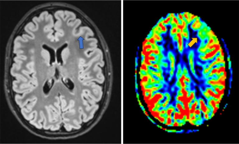
Figure 3.
Left FCD (blue arrow) depicted on FLAIR (left) and ASL (right) which shows a reduction in cerebral blood flow (orange arrow) compared to the surrounding cortex (same patient as Figures 1,2). FCD, focal cortical dysplasia; FLAIR, fluid attenuated inversion recovery; ASL, arterial spin labeling.

