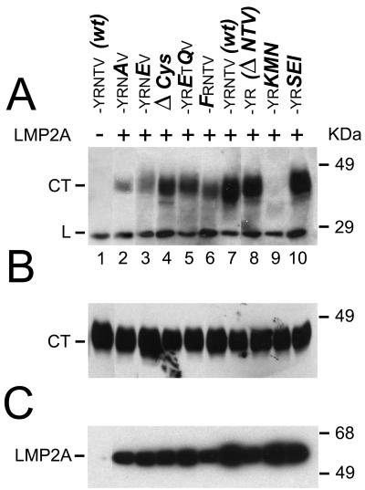
FIG. 1.
Coimmunoprecipitation of CD2-LMP2 CT mutant chimeras with LMP2A. All CD2-LMP2A chimeras were transiently transfected into the stably LMP2A expressing C4 cell line and lysates were immunoprecipitated with the three rat-anti-LMP2A MAbs 8C3, 14B7 and 4E11 coupled to Sepharose beads (see M&M). Captured antigens were electrophoretically separated, transferred to PVDF membranes and probed with the OX34 anti-CD2 MAb (A), to detect the presence of the chimeric proteins in the LMP2A immunoprecipitates. (B) Whole cell lysates from the transfected C4 cells were probed with the OX34 antibody to determine the relative expression of the chimeric CD2 proteins in the transfected C4 cells. The structure of the 27-aa C-terminal sequence in each wt and mutant CD2 chimera, as well as the nomenclature of the mutants, is explained in Table 1. Each lane contains lysate from 105 cells. (C) Filters with the LMP2A immunoprecipitates were probed with a mixture of the three LMP2A-reactive MAbs to demonstrate that the immunoprecipitates from the C4 cells contained LMP2A. Lane 1 in the three panels is lysate from the LMP2A-negative, CD2 LMP2A CT-expressing cell line 3-5, to show that CD2 is not precipitated unspecifically with the LMP2A MAbs.

