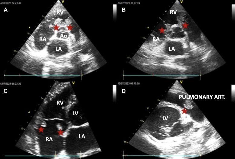
Figure 2.
Echocardiographic sections showing thrombi. (A and B) Short-axis parasternal section passing through the root of the great vessels. (C) Apical four cavities centred on the RV. (D) Modified short-axis parasternal section centred on the trunk of the pulmonary artery. Red stars, thrombi; RA, right atrium; LA, left atrium; RV, right ventricle; LV, left ventricle; Ao, aorta.

