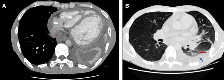
Figure 3.
(A) Axial thoracic scan showing the same thrombi seen on transthoracic echocardiography (red stars) as well as cardiomegaly and dilatation of the left ventricle. (B) Axial thoracic scan showing ground glass (red arrow) opacities and pleural effusion (blue arrow). RA, right atrium; RV, right ventricle; LV, left ventricle.

