
Figure 1.
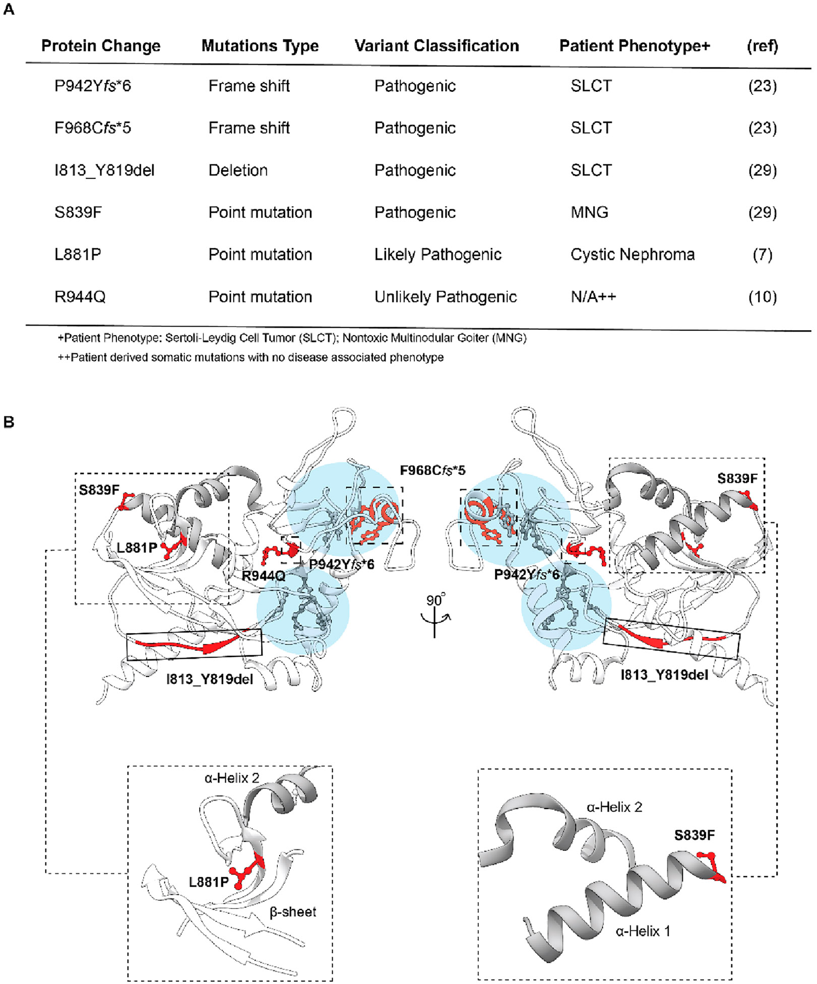
Overview of patient-derived Dicer mutations located within the Platfrom-PAZ domain. (A) Table of patient-derived alterations with corresponding phenotypes. (B) Three-dimensional representation of the Platform-PAZ domain (Protein Data Bank entry 5ZAK) with the location of patient-derived mutations highlighted and close-up views of the secondary structural features surrounding S839 and L881. Residues associated with point mutations are shown as ball and stick models, while frame shift and deletion mutants are grouped by large dashed and solid boxes, respectively. Major RNA-binding pockets are highlighted in light blue with key amino acids colored gray. α-Helices 1 and 2 are colored gray in all models.