
Figure 5.
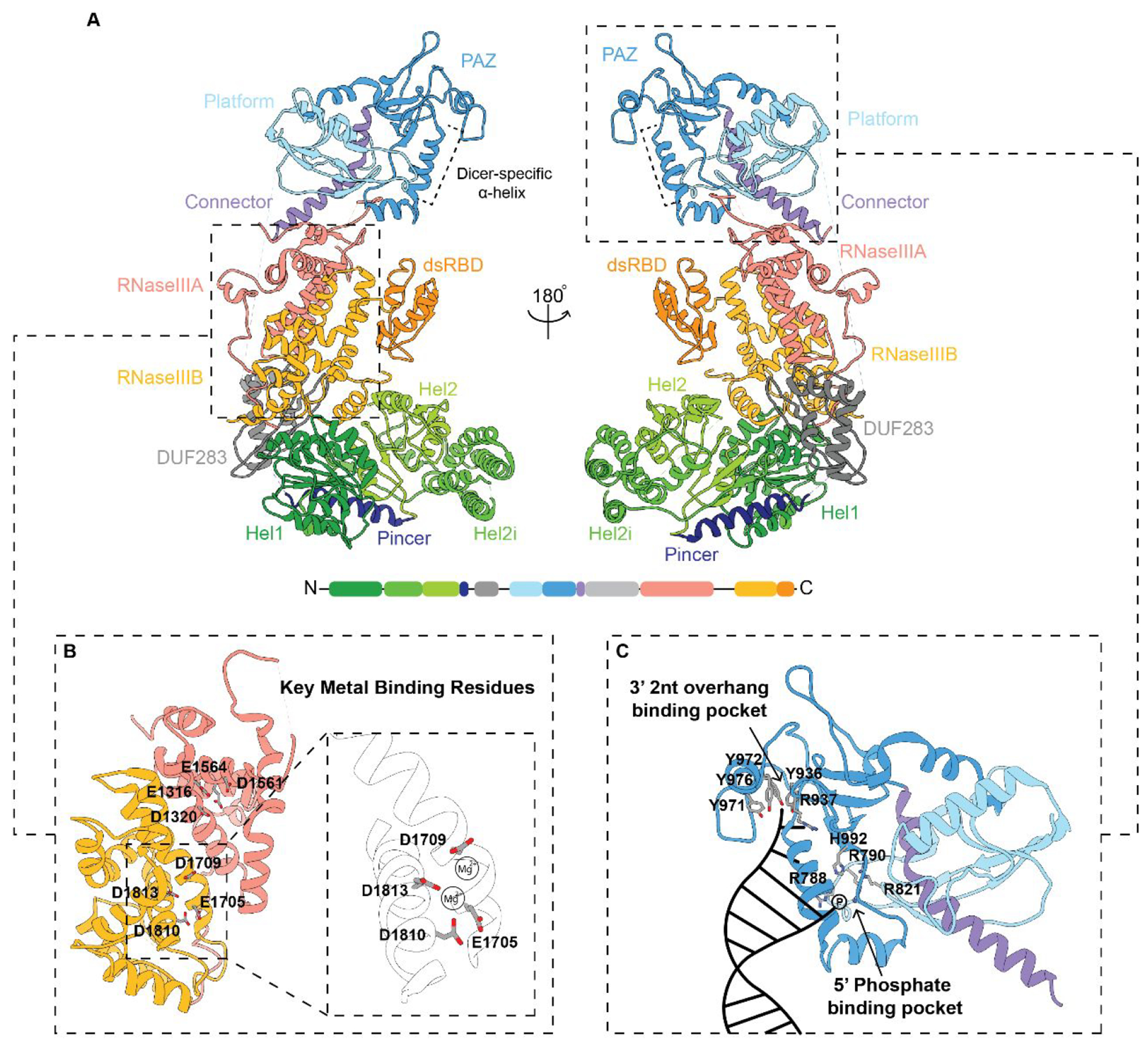
Structural model of human Dicer. (A) Structural overview modeled from a 4.4 Å cryo-EM structure. Domain organization and color code used for labeling the domains from the N- to C-termini are also shown at the bottom. Dashed panels are zoomed-in views of the (B) RNase III processing center and (C) major binding pockets within the Platform-PAZ domains with key amino acids highlighted. A cartoon representation of RNA binding within the 3′ 2 nt overhang and 5′ phosphate binding pockets is depicted in the bottom right panel in (C). (PDB: 5ZAK).