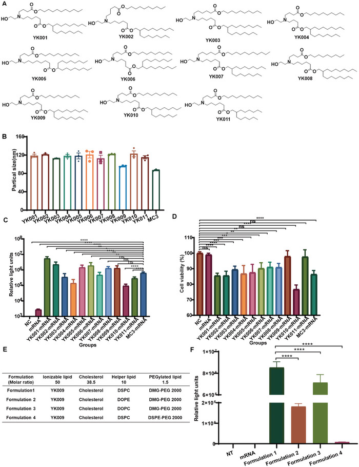
Figure 2.

Characterization and formulation screening of LNPs‐mRNA. A) Chemical structure of 11 ionizable lipids. B) Particle size of LNPs‐mRNA. C) Relative luciferase expression of HEK‐293T cells after incubation with LNPs‐Fluc mRNA for 24 h. D) Cell viability of HEK‐293T cells after incubation with LNPs‐Fluc mRNA for 24 h. E) Formulations of LNPs with different helper phospholipids (1,2‐distearoyl‐sn‐glycero‐3‐phosphocholine (DSPC), 1,2‐dioleoyl‐sn‐glycero‐3‐phosphoethanolamine (DOPE), and 1,2‐dioleoyl‐sn‐glycero‐3‐phosphatidylcholine (DOPC)) and PEGylated lipids (1,2‐dimyristoyl‐rac‐glycero‐3‐methoxypolyethylene glycol 2000 (DMG‐PEG 2000) and 1,2‐distearoyl‐sn‐glycero‐3‐phosphoethanolamine‐N‐[methoxy(polyethylene glycol)‐2000] (DSPE‐PEG 2000)). F) Relative luciferase expression of HEK‐293T cells after transfection with different LNP‐Fluc mRNA formulations. Statistical significance was calculated using one‐way ANOVA (Analysis of Variance), and data are shown as mean ± SEM. (ns: no significant difference, *P < 0.05, **P < 0.01, ***P < 0.001, ****P < 0.0001).
