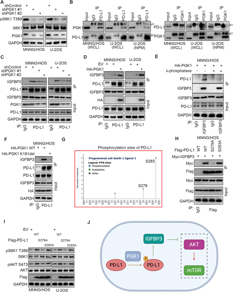
Figure 3.

Hyperphosphorylated PD‐L1 enhances the interaction between PD‐L1 and IGFBP3 in osteosarcoma cells. A) MNNG/HOS and U‐2OS cells were infected with the indicated shRNAs for 72 h. Cells were harvested for western blot analysis. B) The co‐immunoprecipitation was performed in U‐2OS, MNNG/HOS, and NPM U‐2OS by using the IgG, PD‐L1 or PGK1 antibodies. C) MNNG/HOS and U‐2OS cells were infected with the indicated shRNAs for 72 h. Cells were harvested for immunoprecipitation by using the IgG or PD‐L1 antibodies. D) MNNG/HOS and U‐2OS cells were transfected with the indicated plasmids for 48 h. Cells were harvested for immunoprecipitation by using the IgG or PD‐L1 antibodies. E) MNNG/HOS cells were transfected with the indicated plasmids for 48 h. Cells were lysised with RIPA buffer and treated with or without λ‐phosphatase. The whole cell lysates were used for immunoprecipitation by IgG or IGFBP3 antibodies. F) MNNG/HOS cells were transfected with the indicated plasmids for 48 h. Cells were harvested for immunoprecipitation by using the IgG or PD‐L1 antibodies. G) The diagram indicated, the phosphorylation sites of c‐terminus PD‐L1 by analyzing the PhosphoSitePlus® dataset. H) MNNG/HOS cells were transfected with the indicated plasmids for 48 h. Cells were harvested for immunoprecipitation by using the IgG or Flag‐tagged antibodies. I) MNNG/HOS cells were transfected with the indicated plasmids for 48 h. Cells were harvested for western blot analysis. J) A model directing that phosphorylation of PD‐L1 mediated by PGK1 strengthens the interaction between PD‐L1 and IGFBP3 to activate the mTOR signaling pathway in osteosarcoma cells.
