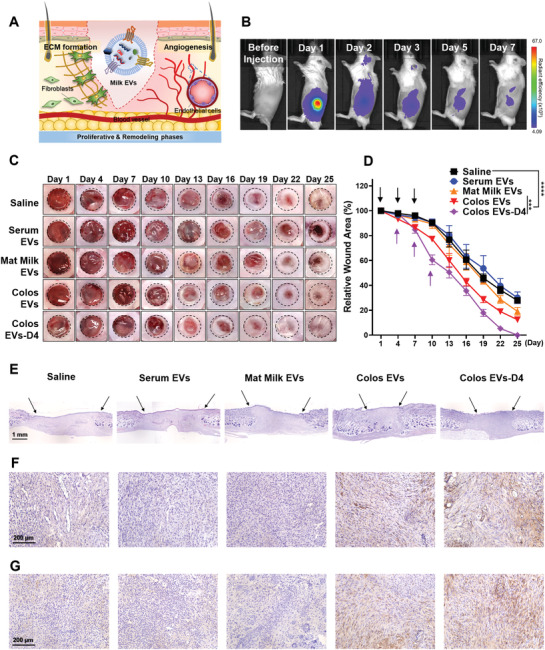
Figure 4.

In vivo therapeutic effect of milk EVs on a cutaneous wound. A) Schematic diagram of milk EVs‐mediated ECM formation and angiogenesis during skin wound healing. B) Images of in vivo biodistribution analysis results obtained for 7 days after subcutaneous injection of Flamma 675 labeled Colos EVs (n = 3). C) Representative images of wound closure after local injection of saline, Serum EVs, Mat Milk EVs, Colos EVs, and Colos EVs‐D4. Mice were injected subcutaneously with PBS or EVs (100 µg per 100 µL PBS) three times at 3‐day intervals. D) Wound size was quantified every 3 days post‐wounding (n = 6). The purple arrows represent the day of injection (days 4, 7, and 10) of Colos EVs‐D4 group. The other groups began EVs treatment from day‐1, as indicated by the black arrows. Data are presented as the mean ± SD (***p < 0.001, ****p < 0.0001 vs saline control; Dunnett's multiple comparisons test). E) H&E staining images of vertically sectioned skin tissues from the excisional wound healing mouse model. The area between the black arrows indicates newly formed dermal tissue. F,G) Representative images of immunostaining for (F) CD31 and (G) elastin proteins in wound tissues from mice treated with saline, Serum EVs, Mat Milk EVs, Colos EVs, and Colos EVs‐D4. All skin tissues were dissected at day 13 post‐wounding and analyzed.
