Figure 5.

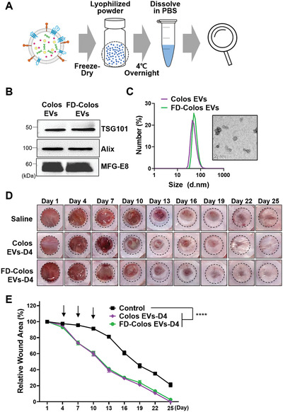
Characterization of lyophilized colostrum‐derived EVs and conservation of in vivo wound healing ability. A) Schematic diagram of the lyophilization procedure. Colostrum EVs were freeze‐dried and stored at −20 °C until use. B) Western blot analysis of EV markers (TSG101, Alix) and MFG‐E8. C) Representative TEM image and size distribution diagram of lyophilized colostrum‐derived EVs. D) Representative images of wound closure after local injection of saline, Colos EVs‐D4, and FD‐Colos EVs‐D4. E) Quantification of wound size at 3‐day intervals post‐wounding (n = 6). The black arrows represent the day of injection (days 4, 7, and 10). Data are presented as mean ± SD (****p < 0.0001 versus saline control; Dunnett's multiple comparisons test).
