
Figure 3.
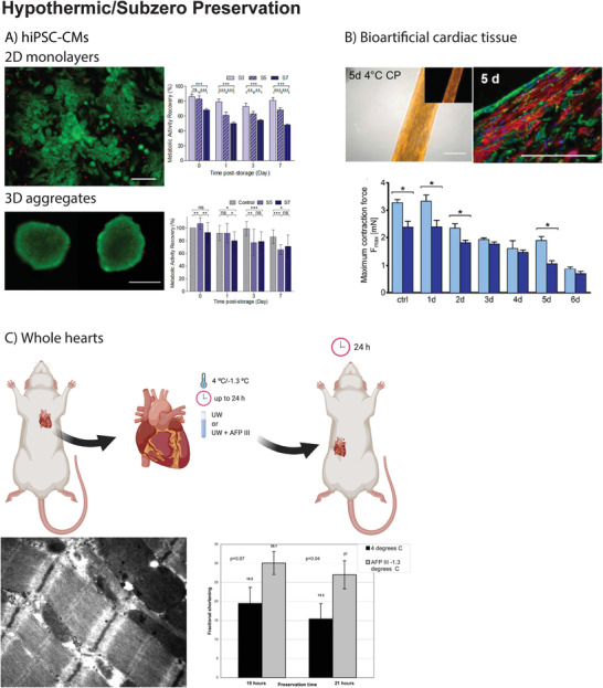
A) In the work by Correia et al., hiPSC‐CMs in 2D monolayers or 3D aggregates were preserved at 4 °C to determine the effect of cell–cell and cell–matrix interactions on preservation success. Cells were preserved in hypothermic conditions for 3, 5, and 7 days (S3, S5, and S7, respectively). After preservation, cells were rewarmed and maintained in culture for up to 7 days. Preservation as 2D monolayers was only feasible up to 3 days, as metabolic activity recovery was significantly compromised for S5 and S7. In contrast, when preserved as 3D aggregates, differences in metabolic activity recovery were less pronounced (e.g., 3D aggregates preserved for 7 days recovered 70% of the metabolic activity 7 days post‐storage). Reproduced with permission.[ 32 ] Copyright 2016, Wiley. B) In the work by Beckman et al., neonatal rat CMs were mixed with 10% Matrigel and 0.9 mg mL−1 rat tail collagen type I in a silicon mold to generate a cardiac construct that was subsequently preserved for 1–7 days at 4 °C. Among the tested preservation solutions, ChillProtec could preserve mitochondrial function after 5 days of preservation (tetramethylrhodamine, methyl ester (TMRM) uptake and staining in active mitochondria, left picture) and cardiomyocyte structure (sarcomeric α‐actinin staining, right picture). In addition, myocardial contraction force was completely preserved after 1 day of preservation at 4 °C but gradually decreased after 5 days of preservation (light blue after 1 day of normothermia, dark blue after 5 days of normothermia). Reproduced with permission.[ 86 ] Copyright 2018, IOP Publishing Ltd. C) In the work by Amir et al., whole rat hearts were preserved ex vivo at −1.3 °C in UW supplemented AFPIII for a maximum period of 24 h. After preservation, hearts were heterotopically transplanted in the abdomen of a recipient rat. 24 h after the transplant, hearts preserved in the presence of AFPIII showed a superior preservation of cardiomyocyte ultrastructure (membrane, nucleus, and mitochondria) and an improved fractional shortening, when compared to hearts preserved in UW at 4 °C. Reproduced with permission.[ 42 ] Copyright 2005, Elsevier. Figure 3C top panel was created with BioRender.com.