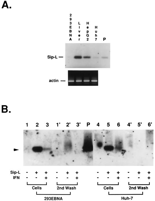
FIG. 7.
Infectivity of HCV in Huh-7 cells stably expressing a higher level of Sip-L mRNA. (A) Sip-L mRNA was detected by RT-PCR and Southern blotting in 293EBNA cells, normal liver tissue, HepG2 cells, and Huh-7 cells. As a control, β-actin mRNA was detected simultaneously. (B) The HCV infection assay was performed with 293EBNA and Huh-7 cells. No HCV RNA can be detected by RT-PCR (arrowhead) in the nontransfected cells (lanes 1 and 4), whereas HCV RNA can be detected in cells expressing Sip-L (lanes 2 and 5). The signals were significantly decreased when 5,000 U of alpha interferon per ml was added to the medium from the 4th day of the infection assay (lanes 3 and 6). The second aliquots of medium used to wash the infected cells were used as contamination controls (lanes 1′ to 3′ and 4′ to 6′).

